Root Canal Treatment (Endodontics)
Upper Left First Molar
Before
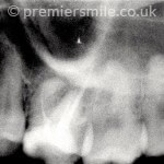
After
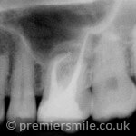
Root canal retreatment of upper left first molar, negotiating the ledged mesial root canal in order to reach the infected apical area.
Lower Right First Molar
Before
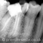
After
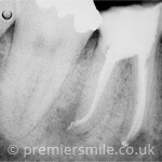
Root canal retreatment of Lower Right First Molar and negotiating the sharp apiral curves.
Upper Right Second Molar and premolar
Before
After
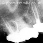
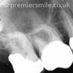
Root canal treatment of Upper Right Second Molar and premolar and retreatment of upper right second premolar. Note the severely curved mesiobuccal root of the UR7.
Apexification
Before
After
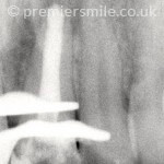
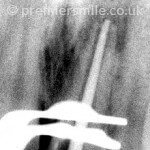
Apexification of the UL1: immature and infected root canal treated and obturated using MTA.
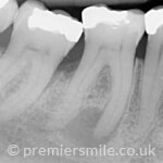
Lower Right First Molar
Before
After
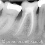
Root canal treatment of lower right first molar with a large periodical lesion (abscess) and complete healing being evident at two years follow up.
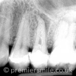
Upper Left Premolar
Before
After
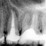
Root canal retreatment of upper left second premolar, including the removal of a broken spiral filler.
Lower Left Second Molar
Before

After
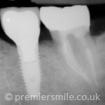
Root canal treatment of lower left second molar. Whilst the LL6 deemed to be unrestorable and had to be extracted. The patient later chose to replace the missing LL6 with an implant supported crown.
Retreated Lower Right Molars
Before
After

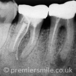
The two year follow up of the retreated lower right first and second molars shows complete healing the periapical lesions and the sinus tract.
Upper First Molars
Before
After

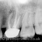
Upper first molars often have two canals which can be located more predictably under the microscope.
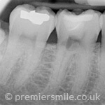
Lower right first molar: Treatment of cervical resorption.
Before
After
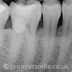
After Root Canal Treatment
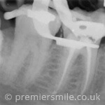
Treatment of cervical resorption was carried out successfully. The pulp retained its vitality for more than 5 years, before it was root treat.


 34, Hockliffe Street, Leighton Buzzard, Bedfordshire, LU7 1HJ


